 Computer Tomography is an advanced imaging modality that utilizes ionizing radiation (X-rays), a series of detectors, sophisticated computers and software to generate cross sectional images or slices. It is advantageous over standard X-ray images because the anatomy of interest can be evaluated as slices without superimposition of other overlying structures. It provides a three-dimensional view of the patient being examined while providing an unparallelled amount of information and insight on the disease processes and complex anatomy.
CT imaging is typically performed before and after intravenous contrast administration under sedation or anesthesia to achieve excellent image detail and contrast resolution.
CT is commonly used for: 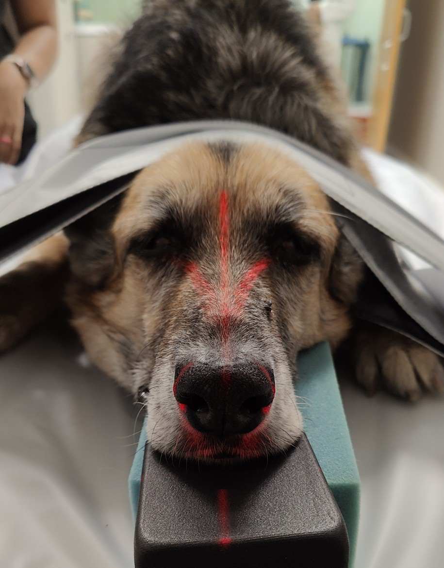
- Diagnosis and staging of cancer
- Surgical planning
- Evaluation of head, neck, chest, abdominal, and limb conditions
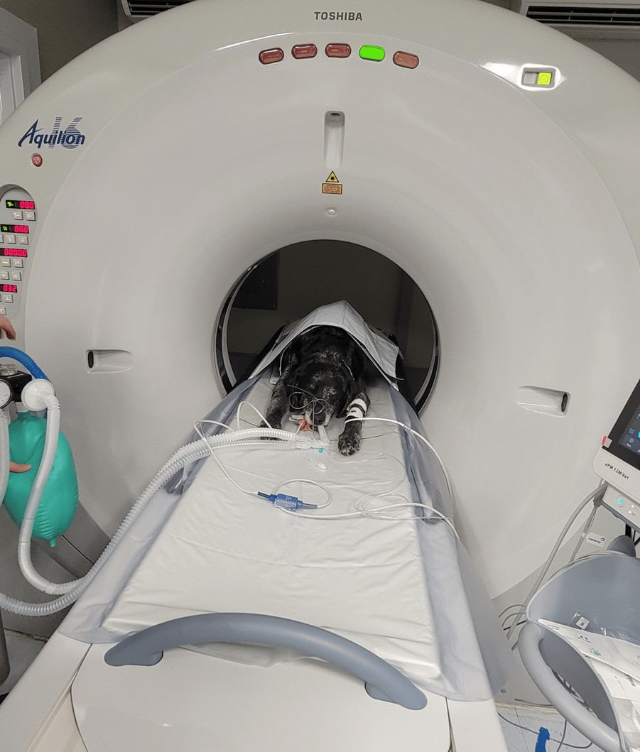
The Diagnostic Imaging service hosts a Toshiba Aquilion 16 slice CT unit that produces excellent studies on both companion and food animal patients that fall within the 450lbs weight capacity.