Radiography
Radiography is often the first line of diagnostic imaging with which the clinician can either make a diagnosis or direct the need for further imaging studies. It is fast, non-invasive, and cost-effective, providing valuable information that can lead to a diagnosis or guide the need for additional imaging studies. Follow-up radiographs may be performed to monitor disease progression or treatment response.
The radiology service performs studies on various species of companion and food animals. Radiographs are routinely used to evaluate:
- Heart and lungs
- Abdominal organs
- Skeletal system
Common conditions diagnosed include heart failure, pneumonia, cancer, foreign bodies, urinary stones, fractures, and hip dysplasia.
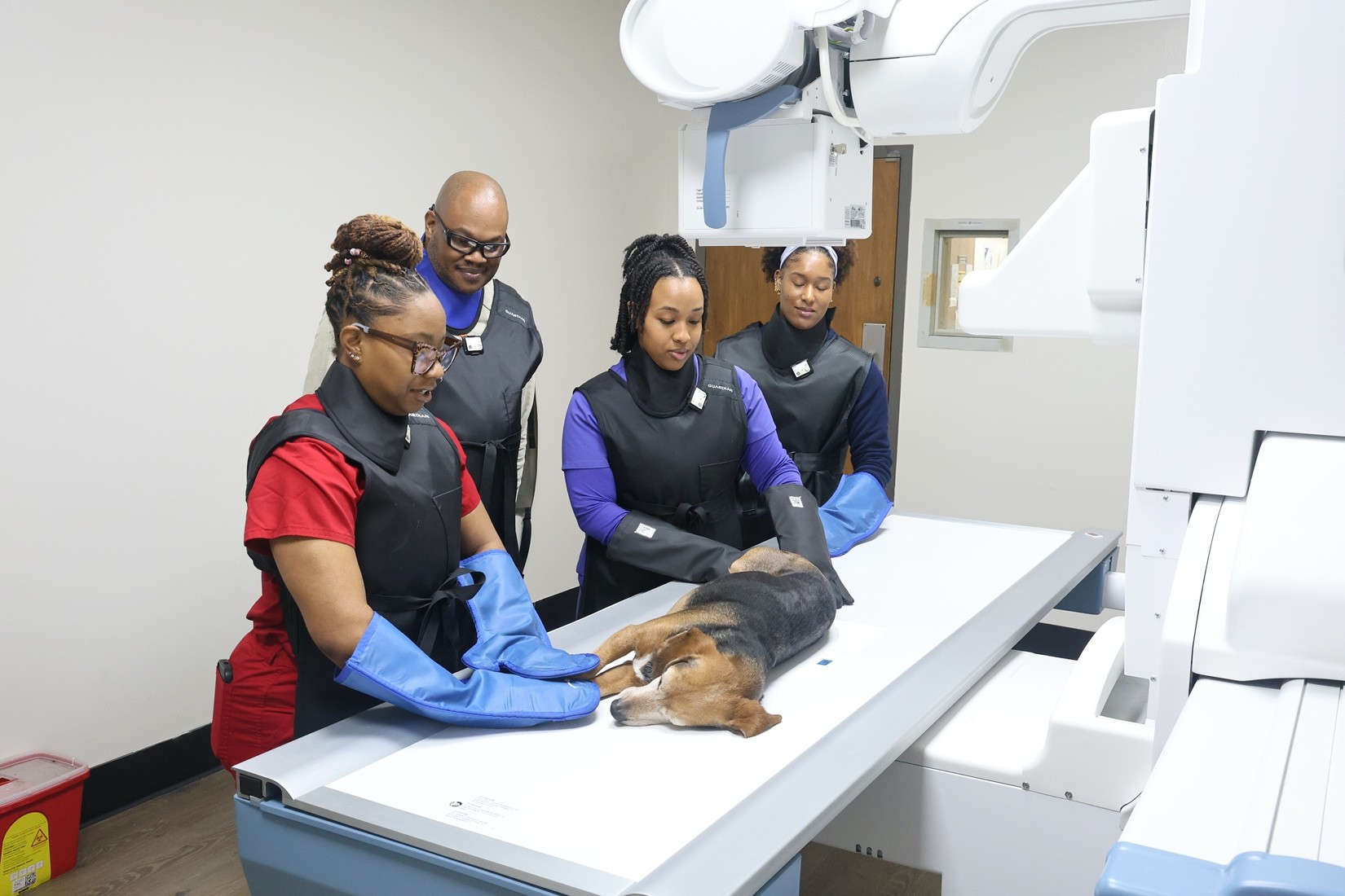
All radiographic studies performed at the Veterinary Teaching Hospital are digital. Digital radiography is the latest iteration of X-ray technology that is now widely available and recognized as having faster processing times, requiring lower radiation exposure and producing higher resolution images. A veterinary technician works closely with the clinical students to perform both routine and specialized exams under direct supervision of an imaging faculty. Advanced procedures such as contrast radiography and positional radiography are also performed under the direction of the imaging clinician. Additionally, Large animal imaging is enhanced by Metron AI software, which integrates artificial intelligence for advanced equine imaging.
All radiographs are reviewed by an in-house, residency-trained radiologist. Images and reports are accessible to clinicians and students throughout the hospital and can be shared electronically with referring veterinarians or specialty services.